Laser Scanning Microscope Magnification

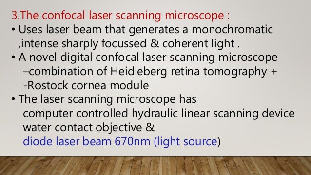
What Is Confocal Laser Scanning Microscopy
Confocal Microscopy Of The Eye
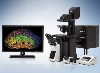
Fv3000 Confocal Laser Scanning Microscope From Olympus Life Science Solutions Get Quote Rfq Price Or Buy
Profile Measuring Laser Microscopes Instruments Used For Roughness Measurements Introduction To Roughness Keyence America

Confocal Laser Scanning Microscopy An Overview Sciencedirect Topics
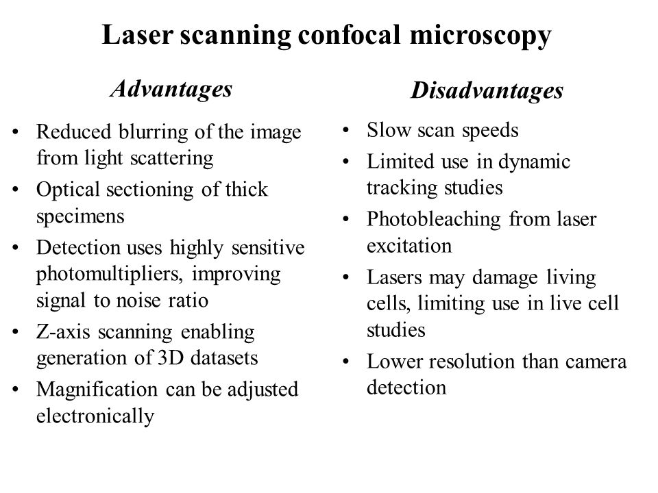
Fluorescence And Confocal Microscopy Ppt Video Online Download

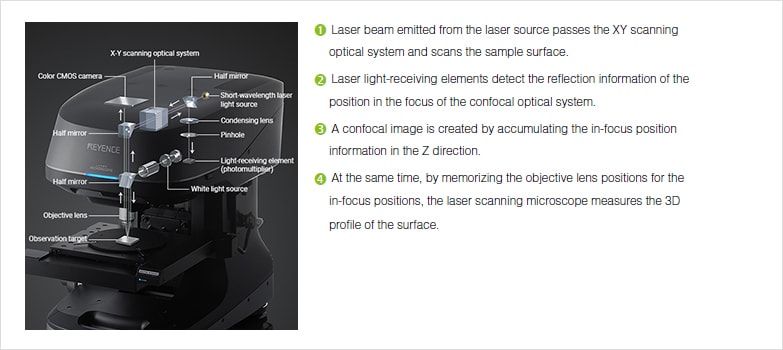
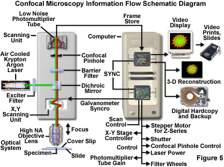
The laser scanning microscope uses a scanning design called beam scanning where the laser image path is scanned in a raster pattern on the surface of the sample.
Laser scanning microscope magnification.
Olympus Fluoview Resource Center Introduction To Confocal Microscopy
Zeiss Microscopy Online Campus Live Cell Imaging Microscopy Techniques

Laser Scanning Microscope 13 Steps With Pictures Instructables
Confocal Microscopy Confocal Microscope Objectives Olympus Life Science

Confocal Laser Scanning Scanning Electron And Transmission Electron Microscopy Investigation Of Enterococcus Faecalis Biofilm Degradation Using Passive And Active Sodium Hypochlorite Irrigation Within A Simulated Root Canal Model Mohmmed 2017
Confocal Microscopy Confocal Microscope Scanning Systems Losungen Von Olympus Fur Den Bereich Life Science
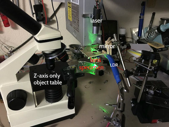
Arduino Blog A Diy Laser Scanning Microscope

Zeiss Microscopy Online Campus Introduction To Spinning Disk Microscopy

A Practical Guide For Fluorescent Confocal Microscopy The Marder Lab

Conducting Steel Plate Surface Texture Topography Analysis With A Laser Scanning Digital Microscope
How Does A Confocal Microscope Work
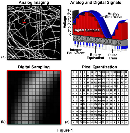
Zeiss Microscopy Online Campus Microscopy Basics Understanding Digital Imaging
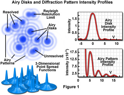
Confocal Microscopy Resolution And Contrast In Confocal Microscopy Olympus Life Science

Confocal Laser Scanning Microscope Labcompare Com

Inverted Zeiss Lsm880 Laser Scanning Confocal Microscope With Airyscan Cell Sciences Imaging Facility Csif

Confocal Laser Scanning Microscope An Overview Sciencedirect Topics


3d Laser Scanning Microscopy Model Vk X200 Keyence Lnnano Lnnano
Https Encrypted Tbn0 Gstatic Com Images Q Tbn 3aand9gcr3fysoxor5w4y0kayjtt5nby84 Yhi3vdxn3rx2 E Usqp Cau
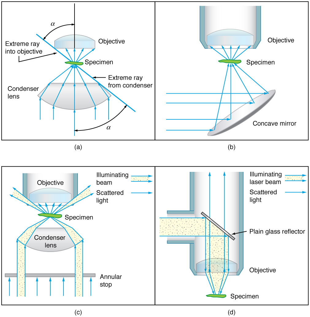
26 4 Microscopes College Physics Openstax

Confocal Microscopy An Overview Sciencedirect Topics
Confocal Microscopy Dinesh

Laser Scanning Microscopes Keyence America
.jpg?rev=33A9)
Fv1200 Olympus Life Science
Source : pinterest.com
