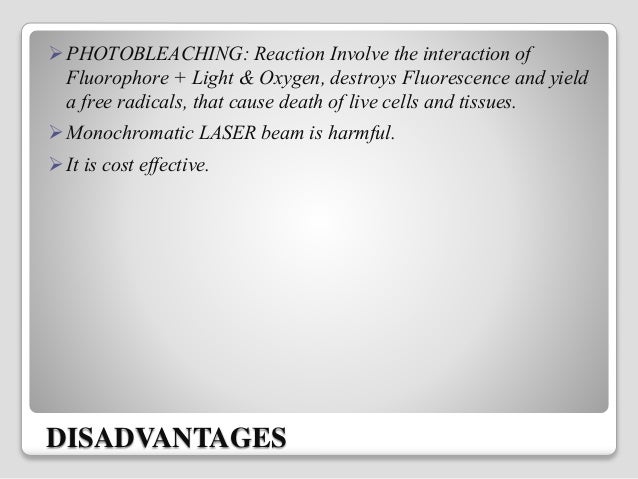
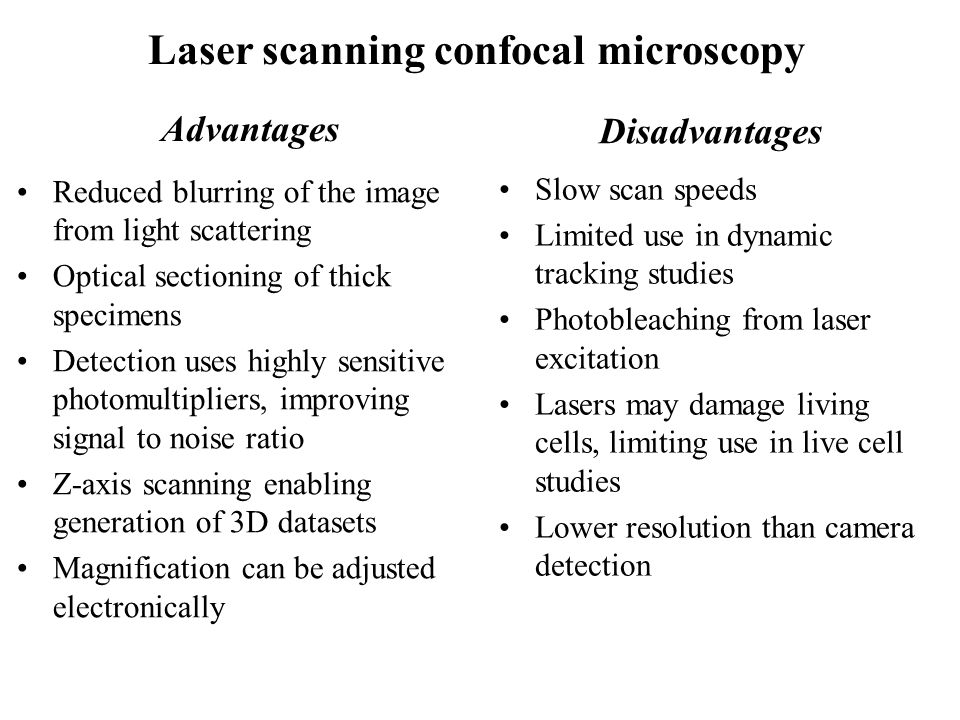
Laser Scanning Microscope Disadvantages

What Are The Limitations Of Confocal Laser Scanning Microscopes Quora

Near Field Scanning Optical Microscopy Advantages And Disadvantages
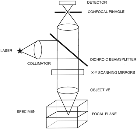
Confocal Laser Scanning Microscopy Clsm

Modern Laser Scanning Confocal Microscopy Bayguinov 2018 Current Protocols In Cytometry Wiley Online Library

The Use Of Laser Scanning Confocal Microscopy Lscm In Materials Science Hovis 2010 Journal Of Microscopy Wiley Online Library

Manual Capsulorhexes Above And Catalys Capsulotomies Below Stained With Trypan Blue Catalys Capsulotomies Exhibit Precisio Cataract Surgery Cataract Laser

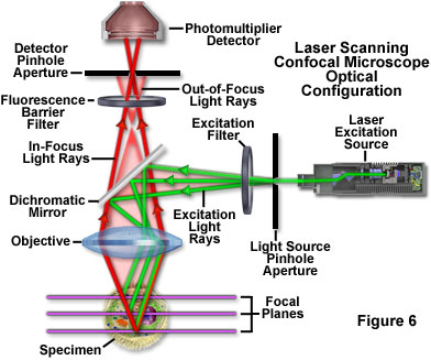
The laser scans across the object and an image is built up pixel by pixel on a screen.
Laser scanning microscope disadvantages.
Zeiss Microscopy Online Campus Live Cell Imaging Microscopy Techniques

World S First White Lasers Demonstrated More Luminous Energy Efficient Than Leds White Lasers Look To Be The Future In Lighting And Li Fi Or Light Based Wir Futuristic Technology Nanotechnology Technology
Spinning Disk Vs Laser Scanning Confocal Microscopes Features Oct 2004 Photonics Spectra
Confocal Microscopy Introduction Olympus Life Science

If You Need Any Optical And Ophthalmic Device Pls Contact Me Email Lisafan Hyvisionstar Com Eye Health Ophthalmic Equipment Optical
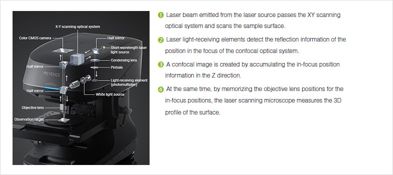
Profile Measuring Laser Microscopes Instruments Used For Roughness Measurements Introduction To Roughness Keyence America

Pdf Confocal Scanning Optical Microscopy And Its Applications For Biological Specimens Semantic Scholar

Tevo Tornado X Axis Tensioner Remix By Gabix Thingiverse Tornado Remix 3d Printing

Python Programming Logo In Stained Glass 105 00 Via Etsy This Would Be A Fun Thing To Put In One Of My Wind Python Programming Language Logo Learn To Code
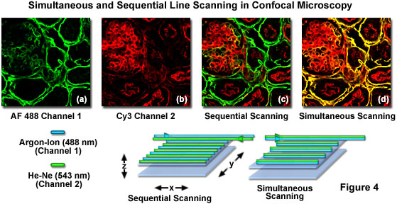
Olympus Fluoview Resource Center Spectral Bleed Through Artifacts In Confocal Microscopy
.jpg)
The Benefits Of Using A Confocal Microscope

Confocal Microscopy An Overview Sciencedirect Topics

Orlas Station Machine Design Cnc Design Industrial Machine
Fluorescence And Confocal Microscopy Ppt Video Online Download
Confocal Laser Scanning Microscopy Springerlink

Tattooremovalproducts Laser Hair Removal Machine Hair Removal Machine Tattoo Removal

What Is Spinning Disk Confocal Microscopy

What Are The Main Differences Between An Sem An Esem An Sem Fib And An S Tem Horiba
Https Encrypted Tbn0 Gstatic Com Images Q Tbn 3aand9gctuan2diibxh Akqhct Gs3wlxe49owmw0 Ho3imvfwzh93skjl Usqp Cau
Review Of Progress In Atomic Force Microscopy Fulltext

Future Directions For Induced Pluripotent Stem Cells Research Stem Cell Research Stem Cells Cell

Fluorophores An Overview Sciencedirect Topics

Confocal Laser Scanning Microscopy An Overview Sciencedirect Topics

Femtosecond Laser Setups For Cell Membrane Poration
Source : pinterest.com
