Laser Scanning Confocal Microscope Maximum Magnification

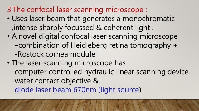
What Is Confocal Laser Scanning Microscopy

Confocal Microscopy Introduction Olympus Life Science

Laser Scanning Confocal Microscopy Lscm Reflexion Mode Images At Two Download Scientific Diagram

Biophotonics Lecture 16 November Fourier Plane Point Object Image F F F F Magnification M 1 Angles Sin Sin Magnification Ppt Download
How Does A Confocal Microscope Work
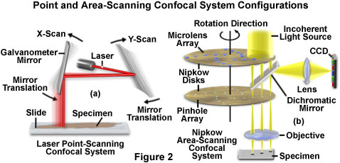
Confocal Microscopy Confocal Microscope Scanning Systems Olympus Life Science

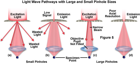
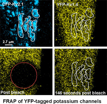
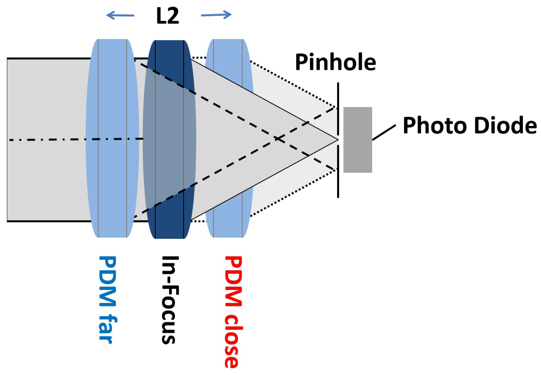
Laser scanning confocal microscopy laser scanning confocal microscopes employ a pair of pinhole apertures to limit the specimen focal plane to a confined volume approximately a micron in size.
Laser scanning confocal microscope maximum magnification.

Confocal Laser Scanning Microscopy An Overview Sciencedirect Topics

A Practical Guide For Fluorescent Confocal Microscopy The Marder Lab

Laser Scanning Confocal Microscope Tailored Tutors

Nikon A1si Laser Scanning Confocal Microscope Washington University Biology Imaging Facility

Introduction To Spinning Disk Confocal Microscopy

33 Laser Scanning Confocal Microscopy And Laser Microdissection Musculoskeletal Key
Zeiss Microscopy Online Campus Introduction To Spinning Disk Microscopy
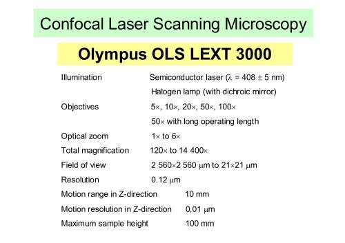
Confocal Laser Scanning Microscopy Olympus Ols Lext 3000
Confocal Microscopes Microscope Imaging Network

Confocal Laser Scanning Microscope An Overview Sciencedirect Topics

Laser Scanning Confocal Microscopy Of Scots Pine Seedlings Colonized By Download Scientific Diagram
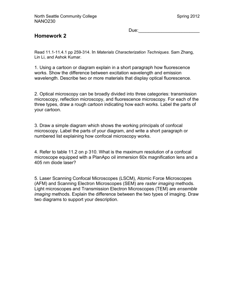
Homework 2 Confocal Microscopy 04 05 12

Confocal Microscopy An Overview Sciencedirect Topics
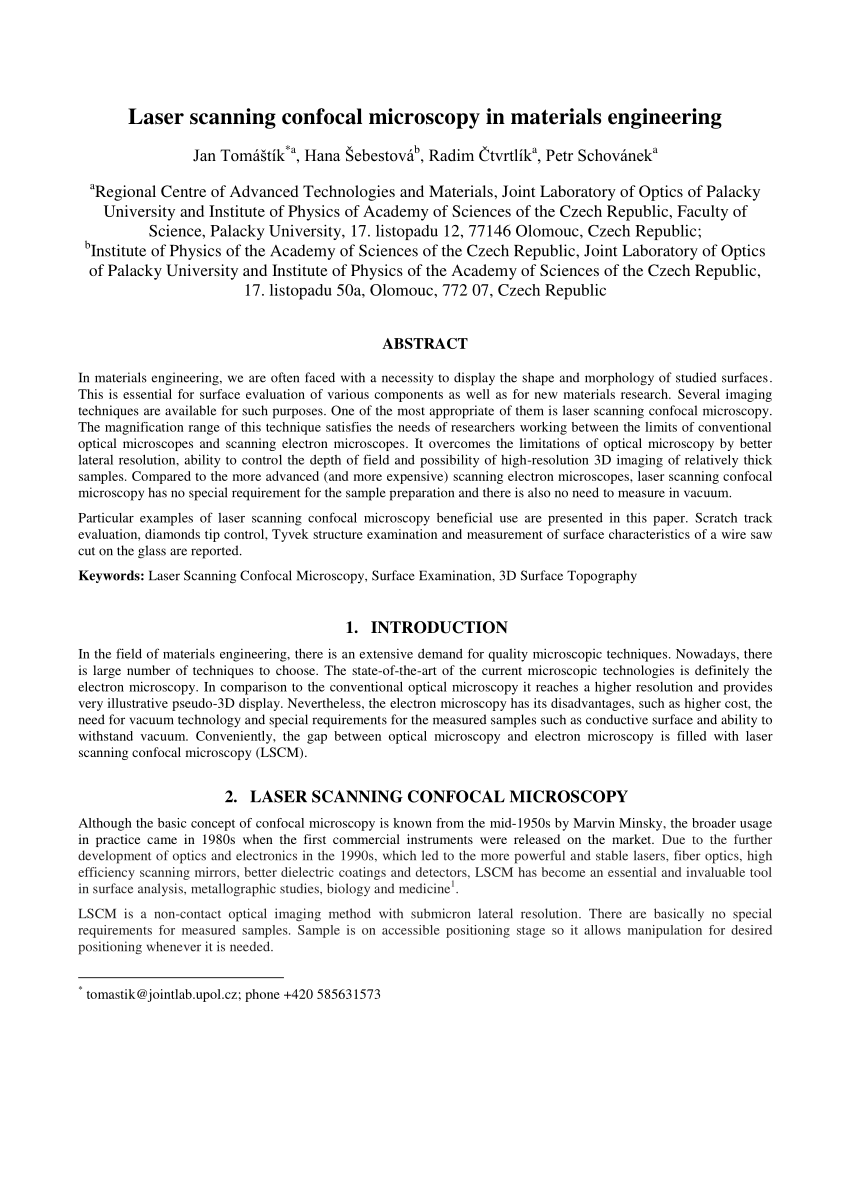
Pdf Laser Scanning Confocal Microscopy In Materials Engineering

Pdf Advanced Microscopy Laser Scanning Confocal Microscopy

Confocal Microscopy For Real Time Detection Of Oral Cavity Neoplasia Clinical Cancer Research

Inverted Zeiss Lsm880 Laser Scanning Confocal Microscope With Airyscan Cell Sciences Imaging Facility Csif

Advances In Bioscience Education Summer Workshop Ppt Video Online Download
Https Encrypted Tbn0 Gstatic Com Images Q Tbn 3aand9gcr3fysoxor5w4y0kayjtt5nby84 Yhi3vdxn3rx2 E Usqp Cau

Laser Scanning Confocal Microscopy History Applications And Related Optical Sectioning Techniques Radiology Key
Fv3000 Confocal Laser Scanning Microscope From Olympus Life Science Solutions Get Quote Rfq Price Or Buy
Applied Sciences Free Full Text Contrast Enhancement For Topographic Imaging In Confocal Laser Scanning Microscopy Html

Olympus Confocal Microscope Lext Ols3100 Tel Aviv University Center For Nanoscience And Nanotechnology
Confocal Microscopy Dinesh
Source : pinterest.com
